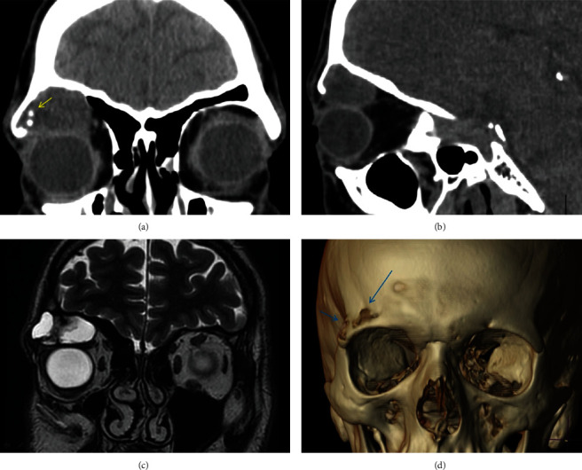
Figure 3.

(a, b) Coronal and sagittal CT images, demonstrating an expansive lesion arising from the roof of the right orbit with soft tissue attenuation. Small foci of mineralization (arrow) into the lesion are presented. The lesion protrudes into the orbit. (c) Coronal T2-weighted MR image shows a well-defined lesion arising from the bone with an extraosseous component. The majority of the tumor has high signal intensity on the T2-weighted image, with low signal areas which represent the solid component of the tumor. (d) Volume-rendered 3D-CT reconstruction images show lytic areas with bone destruction in the roof of the orbit.
