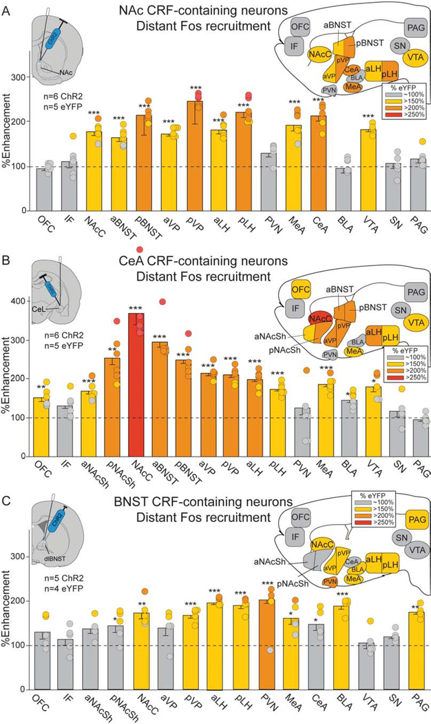
Figure 4. Laser-enhancements in distant Fos expression.
Brain maps show recruitment of distant Fos elevation in mesocorticolimbic structures following CRF-containing neuron ChR2 stimulation in NAc, CeA or BNST (colors denote percent Fos elevation vs. eYFP controls, all two-way unpaired t-tests). A) NAc ChR2 stimulation (n=3 female, n=3 male): NAc core (NAcC), ventral tegmentum (VTA), anterior ventral pallidum (aVP), posterior VP (pVP), anterior lateral hypothalamus (aLH), pLH, medial amygdala (MeA), CeA, aBNST, pBNST. B) CeA ChR2 stimulation (n=3 female, n=3 male): orbitofrontal cortex (OFC), aNAcSh, pNAcSh, NAcC, aVP, pVP, aLH, pLH, MeA, VTA, aBNST, pBNST, and minor increases in basolateral amygdala (BLA; <150%). C) BNST ChR2 stimulation (n=2 female, n=3 male): BLA, periaqueductal gray (PAG), hypothalamic paraventricular nucleus (PVN), NAcC, pVP, aLH, pLH, MeA, and minor increases in pNAcSh (<150%) and CeA (<150%). See Table S1. infralimbic cortex (IF); substantia nigra (SN). Means and SEM reported. *p<0.05,**p<0.01,***p<0.001

