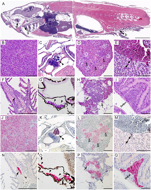
Figure 4. Pathological and immunohistochemical analyses of distant metastases of NB in MYCN transgenic fish with knockout of gas7.
(A-I) H&E-stained sagittal sections of a representative MYCN;gas7mut homo compound fish at 5 months of age. White box in (A) outlines the interrenal gland (IRG), magnified in (B) and (J).
(J–Q) Immunohistochemical analyses with GFP antibody on the sagittal sections of a representative MYCN;gas7mut homo compound fish in magnified views. Disseminated tumor cells were detected in the bone (b, C and K, double black arrowheads), the spleen (D and L, open arrowheads), the liver (E and M, double black arrows), the gill (g, F and N, black arrows), the sclera of the eye (e, G and O, black arrowheads), the pancreas (H and P, open arrows) and the gut (I and Q, double open arrowheads). Scale bars, 2 mm (A) and 50 μm (B–Q).

