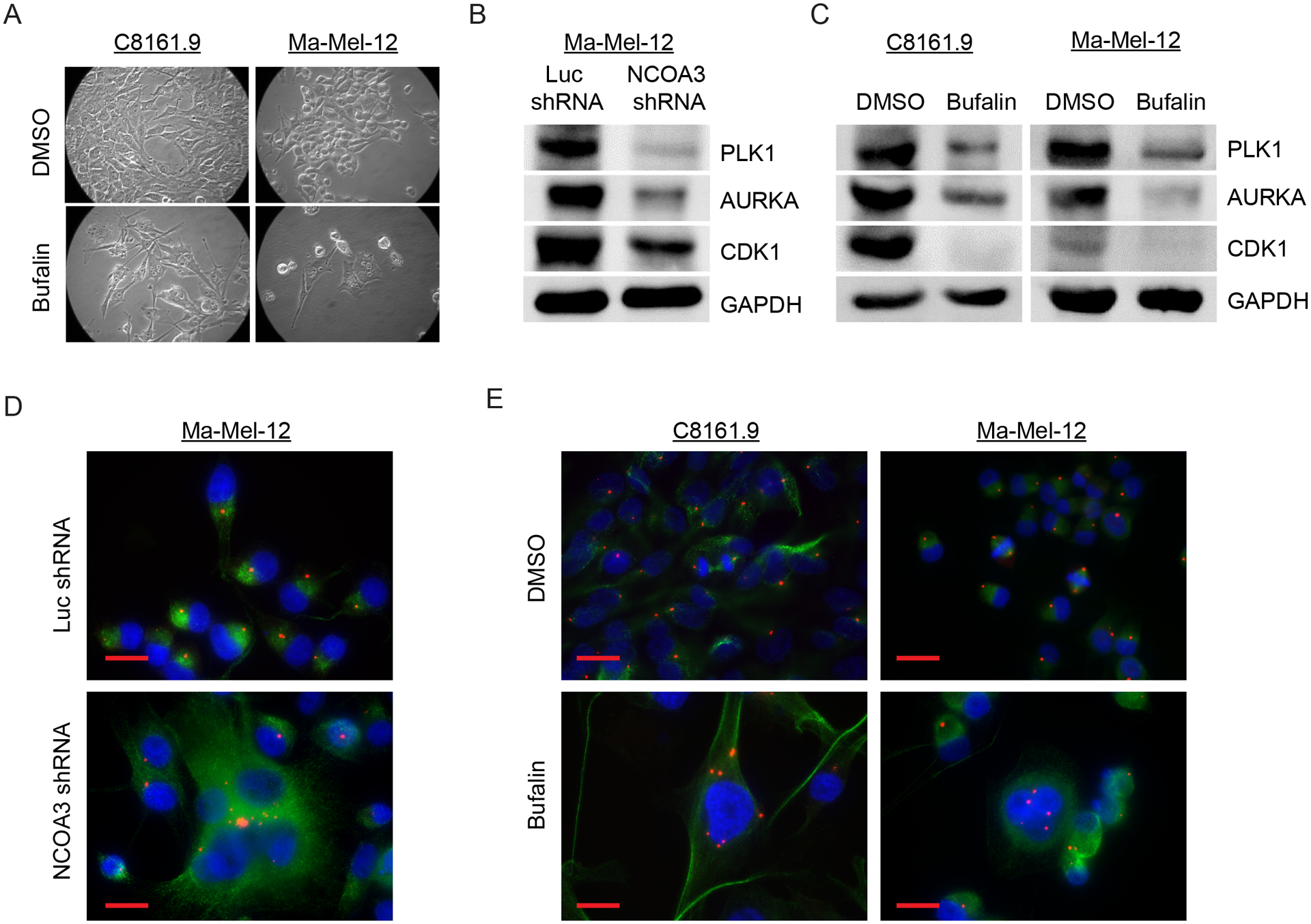
Fig. 4. NCOA3 targeting in melanoma cells results in mitotic catastrophe.

(A) Representative bright field images of C8161.9 and Ma-Mel-12 cells treated with DMSO or bufalin. (B) Western analysis of PLK1, AURKA, CDK1 and GAPDH proteins in Ma-Mel-12 cells stably expressing anti-NCOA3 shRNA or anti-luc shRNA. (C) Western analysis of PLK1, AURKA, CDK1 and GAPDH proteins in C8161.9 and Ma-Mel-12 cells treated with DMSO or bufalin. (D) Representative images of immunofluorescence detecting pericentrin (red) and tubulin (green) in Ma-Mel-12 cells stably expressing anti-NCOA3 shRNA or anti-luc shRNA. (E) Representative images of immunofluorescence detecting pericentrin (red) and tubulin (green) in C8161.9 and Ma-Mel-12 cells treated with DMSO or bufalin. All scale bars are 20 μm.
