
Figure 1.
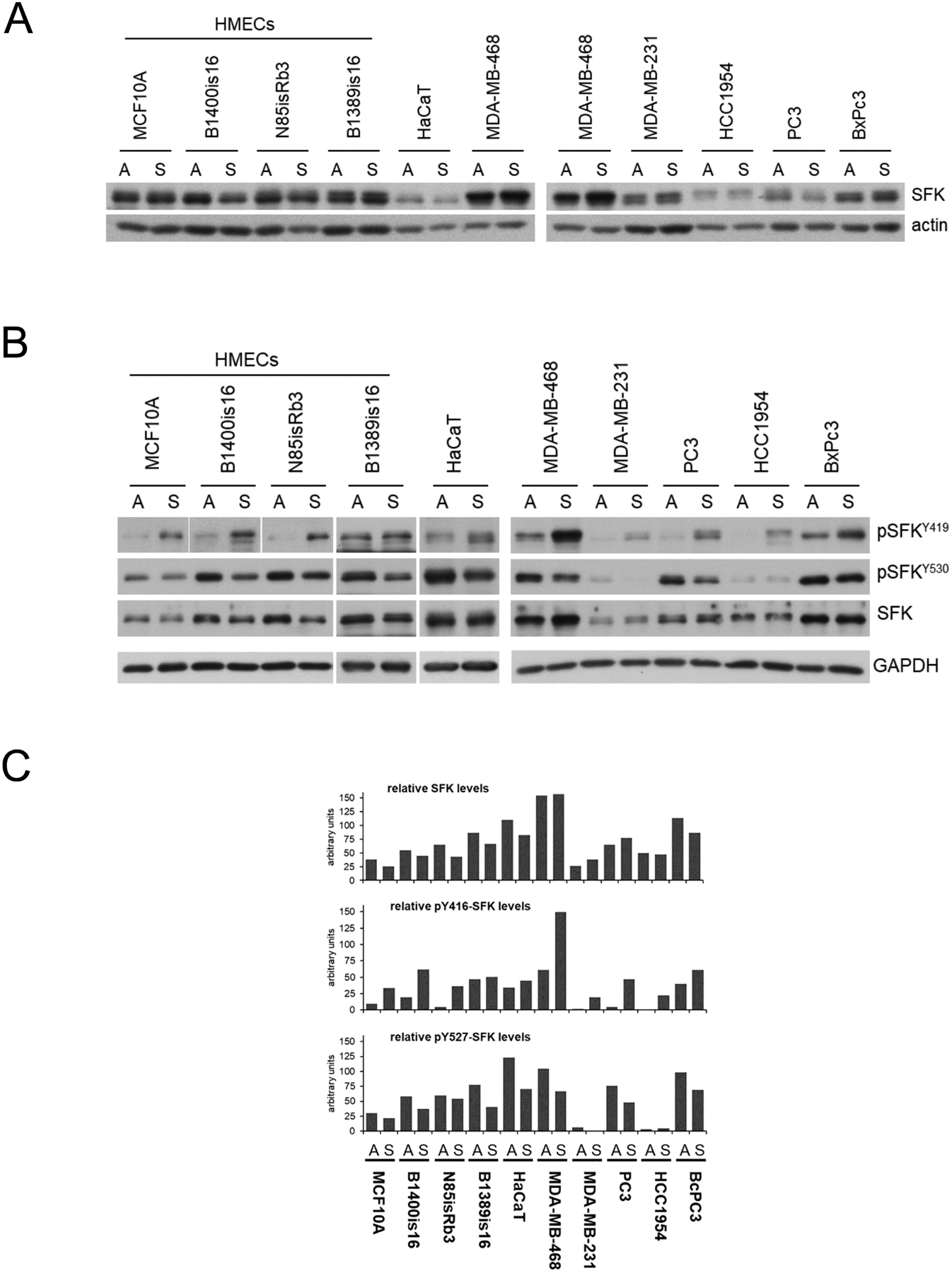
A) Cell lysates from the indicated cancer and non-cancer cell lines were immunoblotted as shown. The SFK antibodies recognize the c-terminal region of this family highly homologous among the family members. B) Cell lysates were immunoblotted against the indicated phospho-specific antibodies marking the highly homologous positive and negative regulatory tyrosines of the SFK family. The Y416 and Y527 residue numbering refers to the commercial names of the antibodies; the actual residue number is different for each SFK family member. C) The immunoblots of part B were quantified, normalized to GAPDH expression, and graphically displayed as shown.