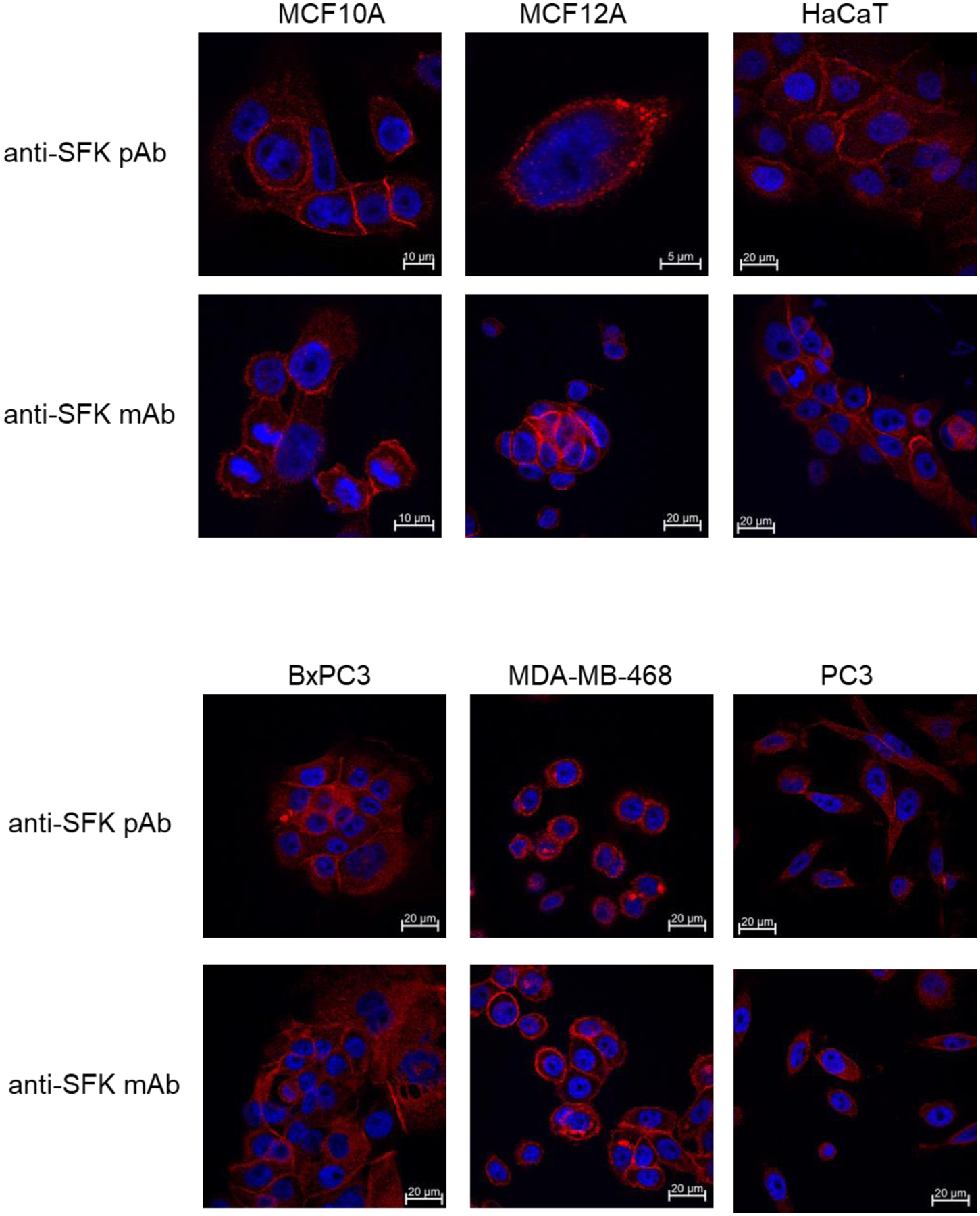
Figure 3.

The indicated cell types were grown on cover slips, fixed in paraformaldehyde and stained using either anti-SFK polyclonal or monoclonal antibodies. These antibodies target the c-terminal homologous region of SFKs and react with Src, Yes, and Fyn. After application of secondary antibodies conjugated with Alexa Fluor 647 the images were acquired using confocal microscopy with the appropriate laser excitation. We used two different antibodies in order to best account for background staining and we used a far-red fluorophore which exhibits much less endogenous fluorescence in most cells. All images were acquired using a 63X objective. Some images are cropped. The scale bars indicate the field size for each individual image. A more comprehensive set of images is provided in Supplementary Figure 2.
