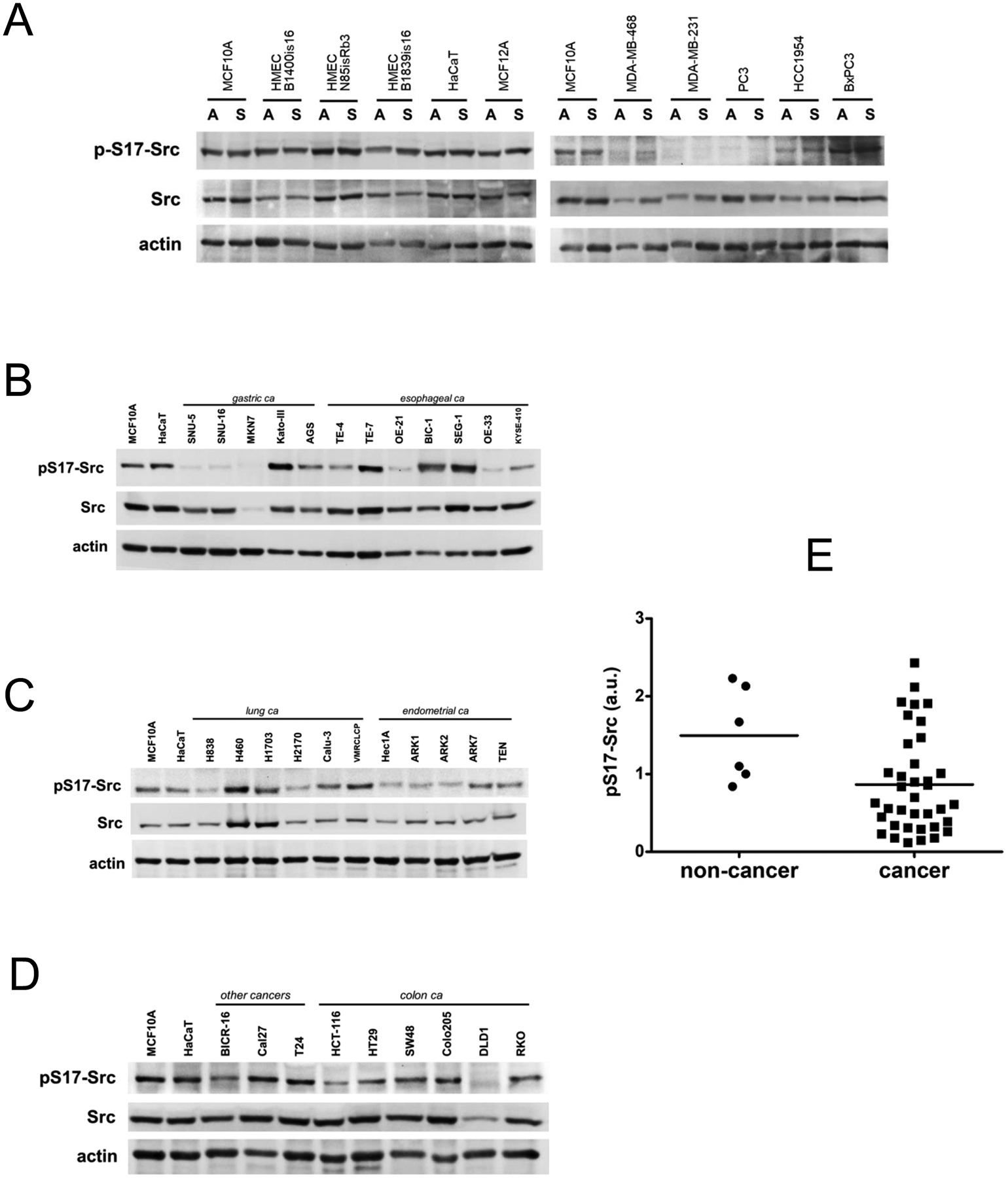
Figure 4.

A) Cell lysates from the indicated cancer and non-cancer cell lines were immunoblotted as shown. Cell lysates were harvested in the adherent state (A) or in the suspended (S) state. S17 is in the unique N-terminal domain of Src and is specific for Src. B-D) Cell lysates from the indicated cell lines in the adherent state were collected and immunoblotted as indicated. The same lysates were loaded in the first two lanes of each gel to allow comparisons across the entire cell panel. E) The level of S17 phosphorylation in each cell line was quantitatively determined by densitometry analysis, normalized to actin, normalized relative to MCF10A on the first lane of each blot, and graphically depicted on the grouped scatter plot.
