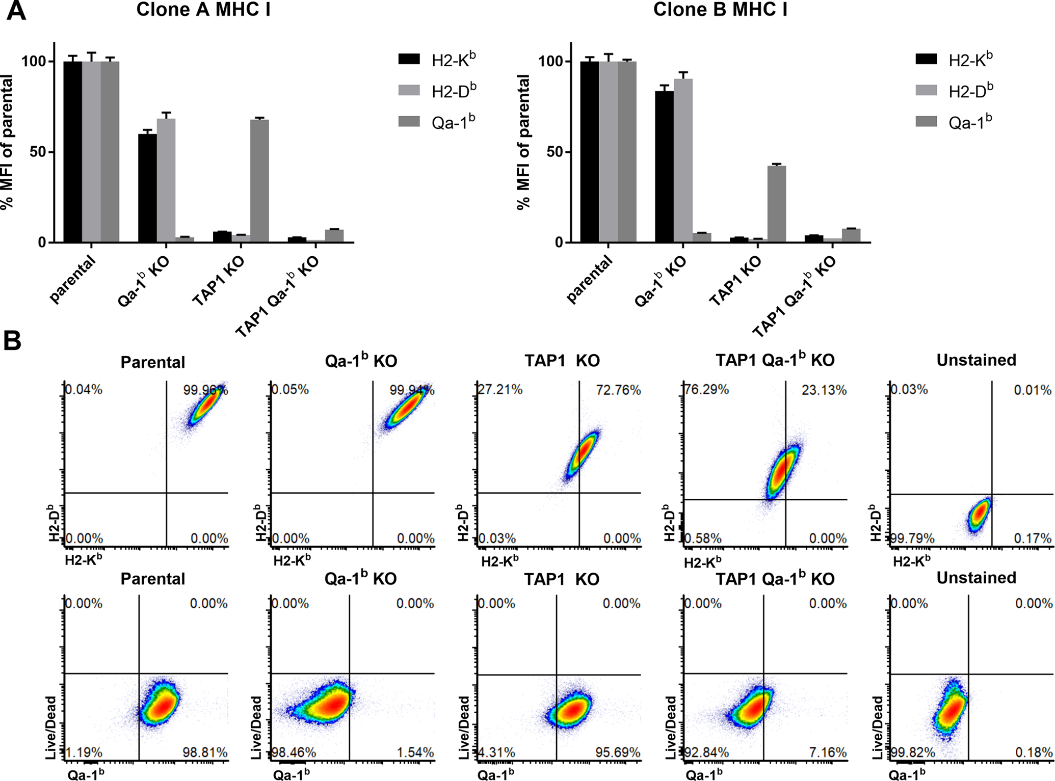
Figure 1. Validation of clonal CRISPR knock-out (KO) cell lines.

Single cell clone phenotypes were confirmed by flow analysis of H2-Kb, H2-Db and Qa-1b cell surface expression. Two sets of single cell clones were selected and used in mouse experiments. Values are shown as mean ± SEM. (A) Flow cytometry of independent clones after 48 h incubation with IFNγ (100 IU/mL). (B) Representative flow cytometry plots.
