Figure 3.

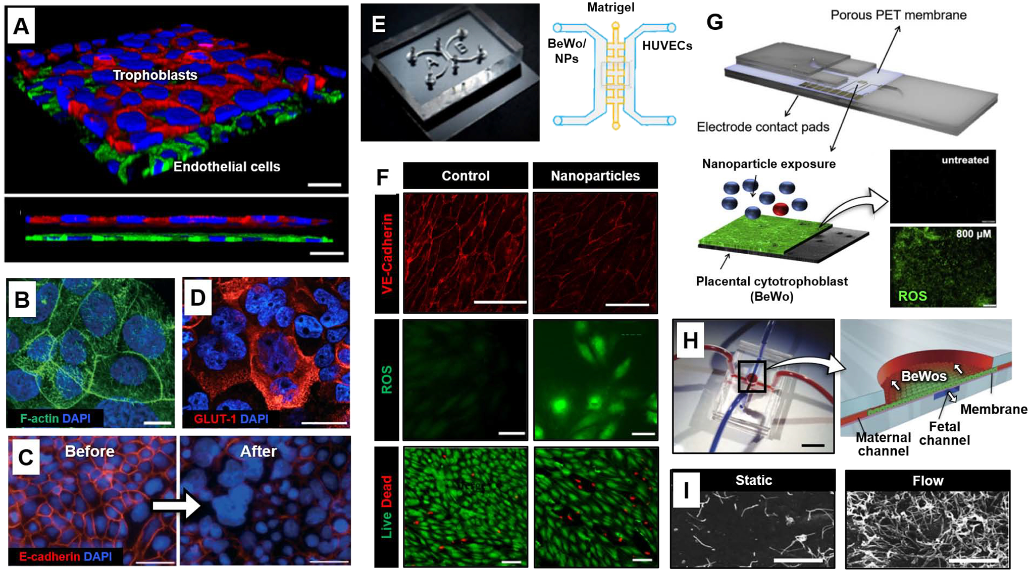
Organ-on-a-chip models of placental barrier function. (A) 3D rendered confocal image of the microengineered placental barrier [56]. BeWo trophoblast and endothelial cells are stained for E-cadherin (red) and VE-cadherin (green), respectively. Nuclei are shown in blue. Scale bars: 30 μm. (B) The surface of BeWo cells cultured under dynamic flow is covered with a dense layer of microvilli (green). Blue shows nuclear staining. Scale bars: 10 μm. (C) E-cadherin (red) and nuclear (blue) staining prior before (left) and after (right) forskolin treatment. Scale bars: 100 μm. (D) Trophoblasts of the microengineered placental barrier express high levels of GLUT-1 (red). Scale bars: 10 μm. (E) Microdevice developed by Yin et al. [58]. Matrigel is seeded in the middle matrix microchannel with BeWo cells and HUVCEs cultured and perfused separately in each side channel. Environmental exposure to nanoparticles (NPs) was modeled by introducing nanoparticles to the maternal (BeWo) side of the chip. (F) HUVEC VE-cadherin expression (red, top), reactive oxygen species (ROS) production (green, middle), and live/dead stain (green/red, bottom) after maternal exposure to titanium dioxide nanoparticles (TiO2-NPs) in the microdevice compared to control. Scale bars: 200, 50, and 100 μm, for VE-cadherin, ROS, and Live/Dead, respectively. (G) Microdevice developed by Schuller et al. [59]. Interdigitated impedance biosensors are located on top of a free-standing porous PET membrane separating BeWo cells and endothelial cells. The rest of the device is fabricated from glass and adhesive layers. BeWo ROS production (green) after 24 hours of exposure to 800 μM zinc oxide (ZnO) particles (bottom) compared to control (top). Scale bar: 200 μm. (H) PDMS device fabricated by Mirua et al. [60]. Red and blue dye were infused into microchannels to visualize the maternal and fetal compartments, respectively (left). Scale bar: 1 cm. Cross-section of device showing the apical maternal channel seeded with BeWo cells and basolateral fetal channel separated by a vitrified collagen membrane (right). The arrows indicate direction of flow in the maternal and fetal channels. (I) Scanning electron micrographs of microvilli protrusions on the apical surface of BeWo cells cultured overnight under static or flow (5 μl/min) conditions. Scale bars: 5 μm. All figures adapted with permissions from original publications.
