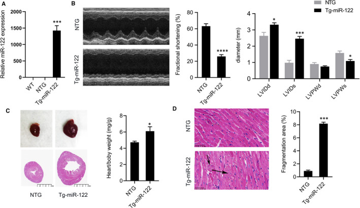
FIGURE 1.

Cardiac overexpression of miR‐122 induced functional deficits consistent with heart failure. (A) Relative expression of miR‐122 in the hearts of Tg‐miR‐122 mice, NTG and wild‐type mice. (B) Representative M‐mode images of Tg‐miR‐122 mice and NTG mice. We quantified the ratio of left ventricular fractional shortening (FS), left ventricular internal diameters at diastole (LVIDd) and systole (LVIDs), and left ventricular wall thickness at diastole (LVPWd) and systole (LVPWs). (C) Haematoxylin/eosin (HE) staining of heart tissue. Scale bar: 2.5mm. Ratios of heart weight to bodyweight (HW/BW) in Tg‐miR‐122 and NTG mice. (D) Representative images of haematoxylin/eosin‐stained heart tissues from Tg‐miR‐122 and NTG mice. Scale bar: 50 μm. N = 5 samples per group. *P < 0.05, ***P < 0.005 for all panels vs NTG mice. An unpaired t test was used to assess significance. Data are means ± SEM
