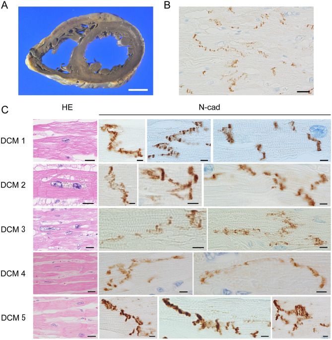
Figure 1.
Pathological and immunohistochemical findings in dilated cardiomyopathy. (A) Macroscopically, we observed severe dilation of the bilateral ventricles. LV wall thickness in the DCM group was relatively uniform, but the wall was thinner in DCM than in the other two groups. (B) ICDs were partially but not clearly observed, and cardiomyocyte units were unclear. (N-cadherin immunostaining; scale bar, 20 µm; original magnification, × 400). (C) Characteristic findings of DCM included cardiomyocyte atrophy, nuclear pleomorphism, and interstitial fibrosis (H–E staining; scale bar, 20 µm; original magnification, × 400). Immunohistochemistry revealed that N-cadherin immunostaining was lower in the DCM group than in the CHF and control groups (N-cadherin immunostaining; scale bar, 5 µm; original magnification, × 1000). ICD width was approximately 2–8 sarcomeres and was significantly elongated in the long-axis direction of the cardiomyocytes. The ICDs were scattered, had a stepwise shape, and were highly curved.

