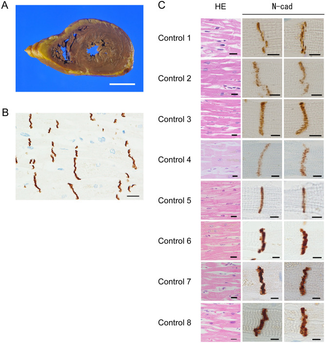
Figure 2.
Pathological and immunohistochemical findings in control cases. (A) Macroscopically, we observed no dilation of the bilateral ventricles. The LV wall had a uniform thickness. (B) ICDs could be clearly observed, and cardiomyocyte units were clear. (N-cadherin immunostaining; scale bar, 20 µm; original magnification, × 400). (C) Histologically, ICDs were observed between cardiomyocytes (H–E staining; scale bar, 20 µm; original magnification, × 400). Each cardiomyocyte could be clearly distinguished. Immunohistochemistry for N-cadherin revealed positive staining at ICDs, and ICDs were thin and flat. (N-cadherin immunostaining; scale bar, 5 µm; original magnification, × 1000). ICD width was within two sarcomeres.

