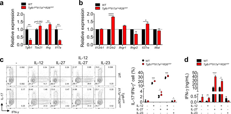
Fig. 3. Th17 cell-specific deletion of TGF-β1 induces conversion of Th17 cells into IFN-γ-producing cells by IL-12 and IL-27.
a, b Tgfb1fl/flIl17aCreR26YFP and WT mice were immunized with MOG35-55 peptide in CFA. Eight to nine days after immunization, lymphocytes isolated from the dLNs were stimulated with MOG35-55 peptide in the presence of IL-23 and an anti-IFN-γ antibody to enrich myelin-reactive Th17 cells. After 5 days, CD4+YFP+ cells were sorted, and relative gene expression was analyzed by qRT-PCR. a Relative expression of Tgfb1, Tbx21, Ifng, and Il17a. b The relative gene expression of cytokine receptors in myelin-reactive Th17 cells was analyzed by qRT-PCR. c, d Th17 cells differentiated from naïve CD4+ T cells were stimulated with anti-CD3ε and anti-CD28 antibodies in the presence or absence of cytokines (IL-12, IL-27, or IL-23) for 3 days and analyzed by flow cytometry and ELISA. c Representative contour plots and quantification of IFN-γ- and/or IL-17-expressing cells in YFP+CD4+ cells. d Quantification of IFN-γ in the supernatant of stimulated Th17 cells after 3 days of culture. Data are representative of three independent experiments. Quantification plots show the mean + SEM (a, b, and d) and ± SD (c); *p < 0.05, **p < 0.01, ***p < 0.001, and ****p < 0.0001. A two-tailed Student’s t-test was performed.

