Figure 1.
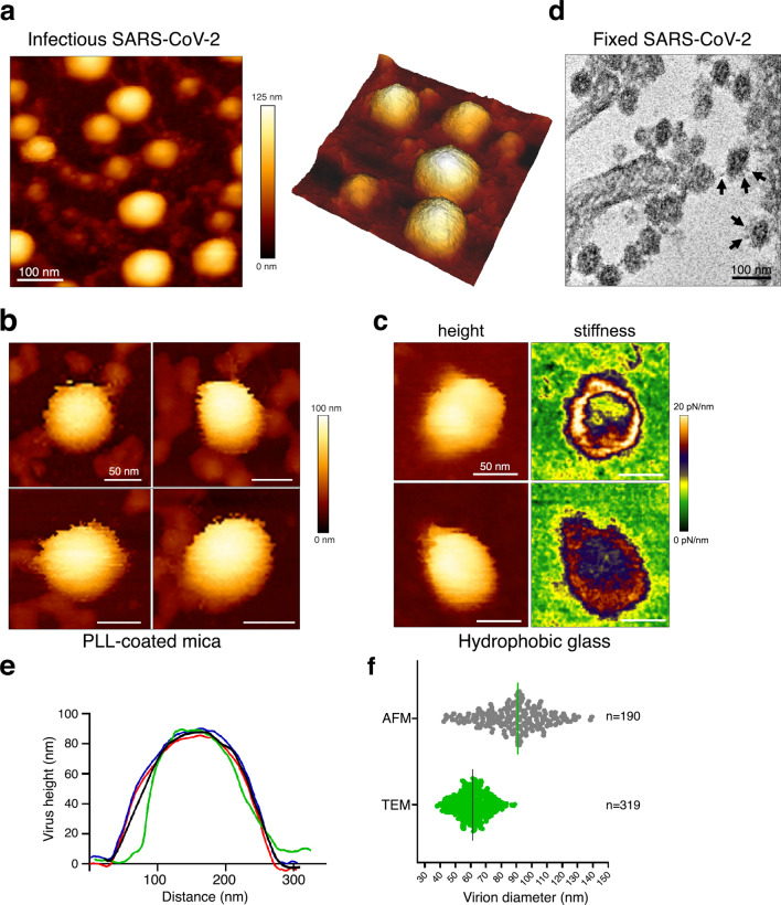
Native infectious SARS-CoV-2 virions imaged by AFM (a) Topographic image and 3D projection of native infectious SARS-CoV-2 virions adsorbed on a poly-l-lysine (PLL)-coated mica surface using quantitative imaging (QI) mode AFM in buffer. (b,c) Zoom-in view of SARS-CoV-2 virions adsorbed on poly-l-lysine-coated mica (b) or glass coverslips coated with an alkyl silane (c) (scale bars: 50 nm). In (c) height and stiffness images acquired in QI mode on individual virions are shown. (d) Example of a TEM image of fixed infected VeroE6 cells producing SARS-CoV-2 that can be seen as spherical particles studded with S trimers (see arrows). (e) Examples of topographical profile plots measured along the horizontal diameter of viral particles. (f) Virion diameter distribution of infectious SARS-CoV2 samples imaged by AFM in liquid (height profile, n = 190 objects) and compared to fixed samples imaged by TEM (n = 319 objects).

