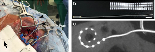
FIGURE 2.

Surgical procedure and intraoperative UC‐MSC‐EV application. (a) Intraoperative image showing the mastoid with the electrode array prepared to be inserted (black bold arrow). Electrocochleography recordings were performed with an extra electrode (black arrow heads). The inner ear catheter (hollow arrow head) is attached to a syringe (asterisk) and contains the EV solution. (b) MED‐EL inner ear catheter with close up of the tip (insert). The tip has three marking spaced 5 mm apart to allow insertion to a predicted depth into the inner ear. Bar equals 1 cm. (c) Postoperative cone beam‐computed tomography showing the intracochlear position of the electrode array of the vesicle‐treated side
