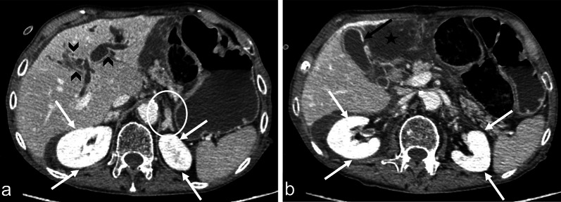
Fig. 7.
Contrast-enhanced CT images in the portal venous phase, in the axial view, shows a 92-year-old male with sepsis of the biliary tract (qSOFA 3) characterised by segmental intrahepatic biliary duct dilatation (a, black arrowheads) and gallbladder leak (b, black arrow) with extrahepatic biloma (b, black star). Note the increased renal parenchymal enhancement bilaterally (a, b, white arrows) and the abnormal adrenal enhancement (a, white circle)

