Figure 7.
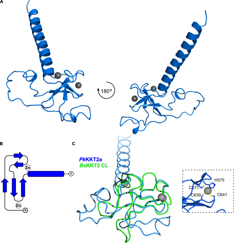
Crystal structure of Perkinsela KKT2a551–679 highlights conservation of CL domain.(A) Cartoon representation of PkKKT2a551–679 in two orientations. Zinc ions are shown in gray spheres. (B) Topology diagram of PkKKT2a551–679 structure. (C) Structure superposition of PkKKT2a551–679 and BsKKT2 CL domain, showing that the core of the structure is conserved. Variations between the two structures are due to sequence insertions within CL and the absence of the C2H2 zinc finger at the C terminus in PkKKT2a551–679. Inset shows a close-up view of zinc ion coordination mediated by the N-terminal residues C571 and H575 and the C-terminal residues C639 and C641.

