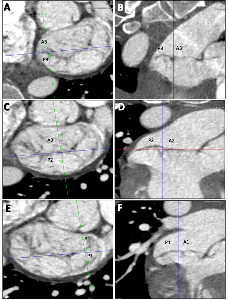
Fig. 1.
Three-chamber views at the level of mitral valve using multiplanar reconstruction to identify prolapse or flail leaflets. Starting with the medial aspect (a, b), the A3 and P3 scallops are identified. In the central portion of the MV (c, d), the A2 and P2 scallops are identified. When scrolling to the most lateral portion of the MV near the LAA (e, f), A1 and P1 are identified. In this example, there is P2 prolapse. MAD is also noted involving all three posterior scallops

