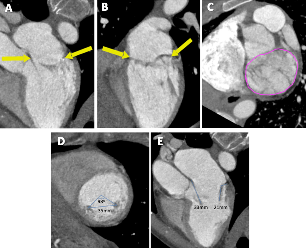
Fig. 2.
Measuring the mitral annulus, papillary muscles, and leaflet length. The boundaries of the mitral annulus are outlined using arrows (a, b), then using planimetry, the MV area is measured in short axis (c). The distance and angle between the two papillary muscles is measured in the short axis (d). Anterior and posterior leaflet lengths are measured in the 3-chamber view in mid-diastole (e)

