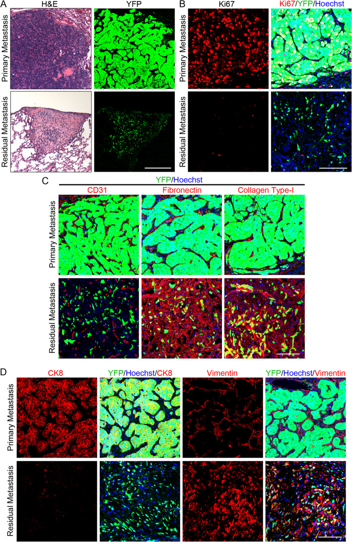
Fig. 5.
Residual metastases exhibit cellular dormancy. a–d Lung metastasis in MTB;TetO-HER2/neu;TTC;rYFP mouse on doxycycline (top) or residual lung metastasis following doxycycline withdrawal (bottom). a H&E-stained sections (left) or YFP fluorescence microscopy (right). b IF for Ki67 (left) or Ki67, YFP and Hoechst 33258 (right). c IF for YFP, Hoechst 33258, and CD31 (left), fibronectin (center) or collagen type-I (right). d IF for CK8 (left), CK8, YFP, and Hoechst 33258 (center-left), vimentin (center-right), or vimentin, YFP, and Hoechst 33258 (right). Scale bars (a) 250 μm and (b–d) 100 μm

