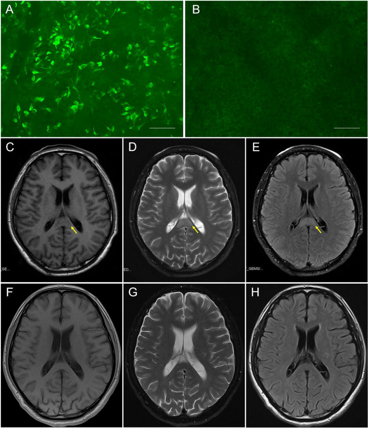
Fig. 1.
Auxiliary examinations of the case with autoimmune GFAP astrocytopathy and RESLES. Antibody test in serum and CSF through indirect immunofluorescence on HEK293 cells (Euroimmun, Lübeck, Germany): A Patient CSF demonstrated binding to the surface of cells expressing GFAP proteins (1:10). B Regions with no specific fluorescence to other proteins on the same slide, analyzed simultaneously, were regarded as negative controls (scale bar: 75 µm). Brain MRI: C–E Initial brain MRI on admission (5 days after onset) exhibited an isolated splenium of the corpus callosum (SCC) lesion (arrows) with hypointensity on T1WI, hyperintensity on T2WI, and on FLAIR. F–H On day 50 after onset (10 days after the second course of immunotherapy), repeat MRI highlighted complete resolution of the SCC lesion

