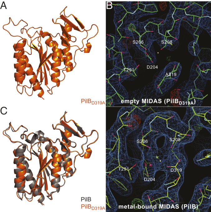
Fig. 5.
3D crystal structure of PilBD319A. (A) Close-up cartoon view of the vWA module in PilBD319A. (B) Comparison of electron density maps in the MIDAS pocket for the PilBD319A (Upper) and PilB (Lower) structures. (C) Superposition of the vWA modules of PilB with bound Mg2+ (gray) and PilBD319A (orange). The two structures superpose with an RMSD of 0.45 Å.

