Fig. 3.
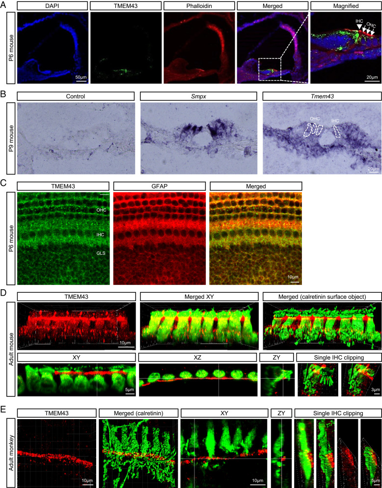
TMEM43 is expressed in the GLSs of mammalian cochlea. (A) Frozen-section images of the mouse cochlea at P6 immunolabeled by anti-TMEM43 (green) and counterstained by phalloidin (red). TMEM43 immunoreactivity is detected mainly at the organ of Corti, and the magnified image shows TMEM43 immunoreactivity at the supporting cells of the organ of Corti. Arrow head, IHCs; arrows, OHCs. (B) In situ hybridization with a control probe, hair cell marker Smpx probe, and Tmem43 probe showing Tmem43 mRNA expression in the Organ of Corti. The dotted lines indicate OHCs and IHCs. (C) Immunostained cochlea tissue with TMEM43 and glial marker GFAP. (D and E) Imaris-reconstructed images of cochlea display TMEM43 expression (red) in GLSs that does not colocalize with hair cells (green) in adult mouse (D) nor adult monkey (E). XY, XZ, and ZY section views and surface object clipping images of a single IHC show that TMEM43 immunoreactivity is found primarily in the apical potion of inner border cell, juxtaposing the upper lateral membrane of calretinin-positive IHC.

