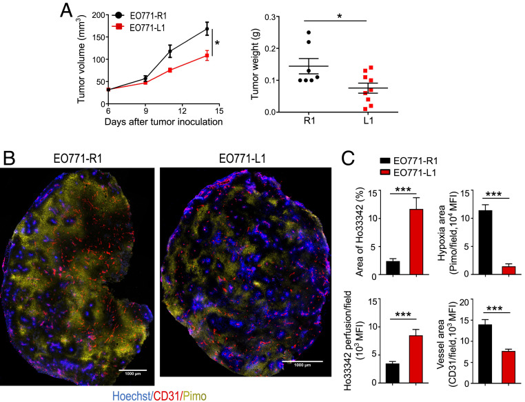
Fig. 1.
Elevated levels of DLL1 in the TME inhibits EO771 breast tumor growth and induces tumor vascular normalization. EO771 murine breast tumor cells overexpressing DLL1 (EO771-L1 or L1) or mock control (EO771-R1 or R1) (2 × 10⁵ cells) were orthotopically inoculated into the mammary fat pad of female C57BL/6 mice. Tumor size was measured every 3 d. The tumor volume was estimated by the formula [(long axis) × (short axis)2 × π/6]. Mice were intravenously injected with 1.2 mg/mouse pimonidazole (Pimo) 25 min and 200 μg/mouse Hoechst 33342 (Ho33342) 5 min before tumor harvest. (A) The tumor growth curves and tumor weight of EO771-R1 and EO771-L1. (B) Representative figures showing Pimo staining and Ho33342 perfusion. (C) The statistical analysis of tumor vessel perfusion (Ho33342), tumor hypoxia (Pimo), and vessel density (CD31) in EO771-R1 and EO771-L1 breast tumors. (Scale bars, 1,000 μm.) Ho33342 (blue), Hoechst 33342 perfused tumor area; CD31 (red), endothelial cells; Pimo (yellow), hypoxic tumor area; MFI, mean fluorescence intensity. Significance was determined by unpaired two-tailed Student’s t tests. Data are from one experiment representative of three (in A, n = 7 to 9 mice per group) or two (in B and C, n = 8 to 10 mice per group) independent experiments with similar results. All data are presented as means ± SEM, *P < 0.05, ***P < 0.001.

