Fig. 2.
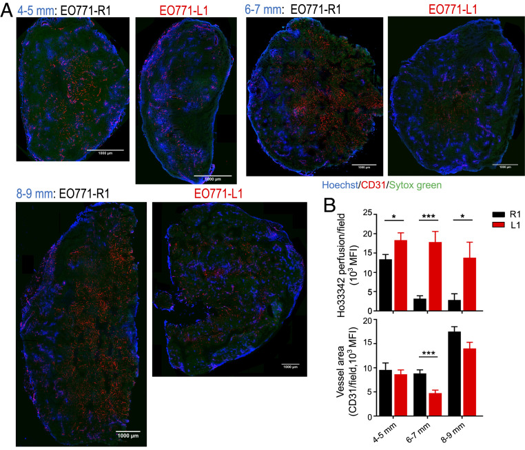
DLL1 elevation in the TME induces long-term tumor vascular normalization. EO771-R1 and EO771-L1 breast tumors were prepared as described in Fig. 1. Tumor tissues were harvested when their sizes reached 4 to 5, 6 to 7, and 8 to 9 mm in diameter, respectively. Vessel perfusion over the entire cross-section of tumor tissues was assessed by confocal microscopy. (A) Representative whole tumor tissue perfusion images. (Scale bars, 1,000 μm.) (B) Vessel perfusion and vessel density in indicated sizes of EO771-R1 and EO771-L1 tumors. Ho33342 (blue), Hoechst 33342 perfused area; CD31 (red), endothelial cells; and Sytox Green (green), counterstained for tumor tissue. Significance was determined by unpaired two-tailed Student’s t tests. Each group had 8 to 10 mice. *P < 0.05, ***P < 0.001.

