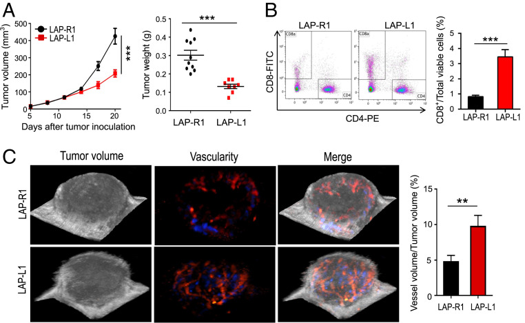
Fig. 4.
Increased levels of DLL1 in the TME inhibits LAP0297 lung tumor growth and induces tumor vascular normalization. LAP0297 murine lung cancer cells overexpressing DLL1 (LAP-L1) or mock control (LAP-R1) (2 × 10⁵ cells) were inoculated subcutaneously in the right flank of female FVB mice. Tumor size was recorded and analyzed as described in Fig. 1. Murine ultrasonographic imaging was conducted to measure global tumor vessel perfusion by using a 3D imaging motor and color Doppler mode. (A) The tumor growth curves and tumor weight of LAP-R1 and LAP-L1. (B) The proportions of tumor-infiltrating CD8+ and CD4+ T cells were analyzed by flow cytometry. (C) The percentages of global tumor blood volume in 3D tumor volume were analyzed. The blue and red colors represent different blood flow directions. The significance was determined by unpaired two-tailed Student’s t tests. Data are from one experiment representative of three independent experiments with similar results (n = 8 to 10 mice per group). All data are presented as means ± SEM, **P < 0.01, ***P < 0.001.

