Fig. 3.
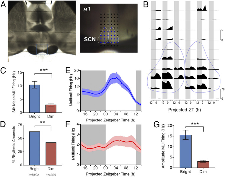
Impact of daytime irradiance on SCN activity. (A) Image of a brain slice of R. pumilio placed over the electrode terminals of a 60-channel array to record neuronal electrical activity in the SCN. The recording site is shown in close up on Right (a1), with dotted white lines delineating the SCN area and electrodes designated as within the SCN shown in blue. (B) Neuronal MUA recorded at the array of electrodes for the preparation shown in A as a function of projected ZT for a 24-h recording epoch. Spontaneous activity within the SCN (bounded by dotted lines) is higher than surrounding hypothalamus and plotted on a different scale (scale bars to right). (C) Time-averaged firing rate (MUA) across 24-h recording epochs from all channels covering SCN harvested from bright (n = 92 channels from four slices) versus dim (n = 99 channels from four slices) conditions. (D) Percentage of channels within the SCN classified as rhythmic for each experimental condition (bright versus dim, **P < 0.01; Fisher’s exact test, data from four slices per condition). (E and F) Multiunit firing rate as a function of projected ZT for rhythmic recording sites within the SCN from animals maintained under bright (E) and dim (F) daytime conditions (bright, n = 58; dim, n = 42 channels; from a total of four animals per condition). (G) Peak-trough amplitude in the multiunit firing rhythm was significantly lower under dimmer lights. (***P < 0.001, Mann–Whitney U test, bright, n = 58; dim, n = 42 channels). Gray background indicates the projected period of lights off (ZT12 to ZT24). Data are expressed as mean ± SEM.

