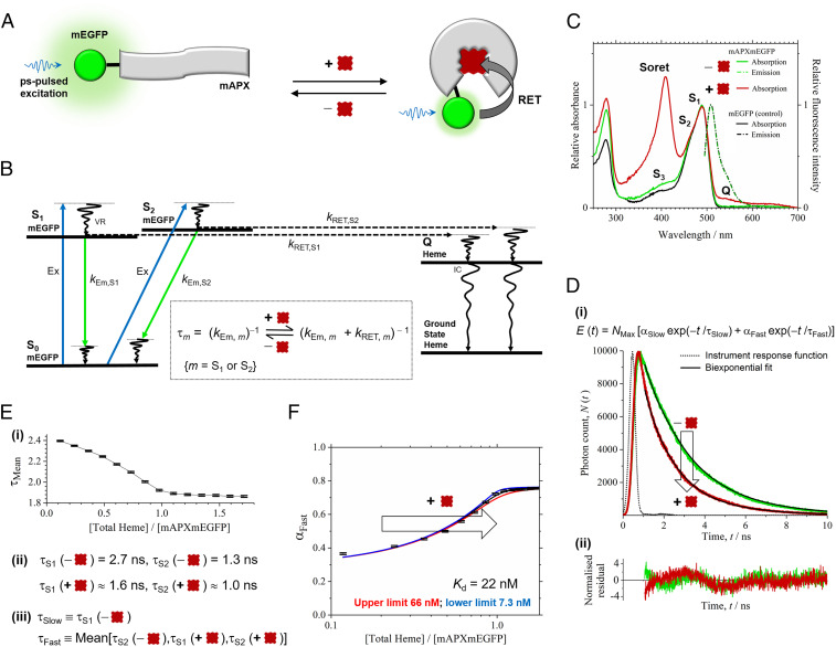
Fig. 1.
Heme sensing in cells. (A) The sensor is mAPX (gray) conjugated to mEGFP (green). Binding of heme (dark red) to mAPX results in RET from photoexcited mEGFP, with concomitant reduction in emission lifetime. (B) The principle of the sensor operation is based on different decay pathways from excited states of mEGFP in the presence and absence of heme. Excitation (Ex, blue) of mEGFP from the ground state, S0, to excited-vibronic states, S1 and S2, is followed by vibrational relaxation (VR). Decay pathways from S1 and S2 are either (A) fluorescence emission (green) with rate constants, kEm,S1 and kEm,S2, respectively, or (B) RET to an electronic-excited state of heme (c.f. mEGFP emission and heme Q band for overlap) with rate constants, kRET,S1 and kRET,S2, respectively, followed by VR and internal conversion (IC) to the ground state of heme. (Inset) Equations for fluorescence lifetimes, τS1 and τS2, in the presence and absence of heme. (C) Absorption and fluorescence spectra for mAPXmEGFP and mEGFP (λEx, 488 nm). Partially resolved bands (centered at 490 nm) for mEGFP are assigned to S1 and S2; a further weak absorption band for mEGFP (S3) and, on addition of heme, Soret and Q bands (408 and 541 nm) are observed. (D) (i) Time-correlated single-photon counting, N (t), from apo- (green) and holo-mAPXmEGFP (red) (λEx, 475 nm; λEm, 510 nm; 37 °C) fitted to a biexponential-decay function, E(t), with time constants of 2.7 (τSlow) and 1.3 ns (τFast). (ii) Normalized residuals for fitting to the decay profiles, (N(t) − E(t)) / √E(t). χ2 values were between 0.8 and 1.7 for biexponential fitting to the reported decay data. Significant improvement in χ2 cannot be achieved by inclusion of >2 decay terms. χ 2 = (1 / h) × Σ (N − E) 2/E {h time bins}. (E) (i) Sequential additions of heme to apo-mAPXmEGFP lead to a gradual increase in the amplitude of the fast (αFast) relative to the slow (αSlow) component and a decrease in the mean lifetime, τMean. (ii) τS1 and τS2 for apo-mAPXmEGFP are the same as the optimized values of τSlow and τFast from E(t). Both τS1 and τS2 are reduced in holo-mAPXmEGFP (see B, Inset), and estimates have been made for these values by assuming that kRET,S1 = kRET,S2 (SI Appendix). (iii) The biexponential model, E(t), for mixtures of apo- and holo- mAPXmEGFP has a slow-decay component, τSlow, equal to τS1 (apo) and a fast-decay component, τFast, equal to the mean of τS2 (apo), τS1 (holo), and τS2 (holo). (F) A theoretical single-site binding model fitted to the amplitude, αFast, in decay profiles obtained by sequential additions of heme to apo-mAPXmEGFP at 37 °C. The estimation of error bars in E and F is described in SI Appendix, section 2.

