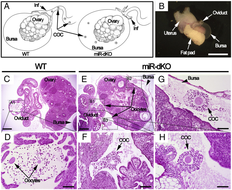
Fig. 3.
Ovulated oocytes are trapped inside the ovarian bursa cavity in miR-dKO female mice, as revealed by whole-mount histology analyses. (A) Schematics showing our hypothesis; that is, the ovulated COCs are picked up by fimbriae of the infundibulum and further transported to ampulla for fertilization in WT oviducts (Left), whereas the ovulated COCs fail to be picked up by fimbriae of the infundibulum in miR-dKO female mice and consequently fall into the ovarian bursa cavity (Right). (B) Gross morphology of the female reproductive system ∼3 h after ovulation, including the oviduct, bursa, ovary, fat pad, and uterus. (Scale bar, 5 mm.) (C) A cross-section showing histology of the ovary, oviducts, and bursa. (Scale bar, 0.5 mm.) (D) Digital enlargement of the ampulla region of the oviduct (framed in C), where two oocytes (arrows) are present. (Scale bar, 0.1 mm.) (E) A cross-section showing that the ovulated oocytes are trapped inside the bursa cavity in miR-dKO mice. (Scale bar, 0.5 mm.) (F–H) Digital magnification of three areas (framed in E), where individual COCs are present inside the bursa. (Scale bar, 0.1 mm.) Both WT and miR-dKO mice in this experiment were primed with pregnant mare’s serum gonadotropin and hCG, and the samples were collected 15 h after hCG injection. COC, cumulus-oocyte complex; Inf, infundibulum. n = 4 for each genotype.

