. 2020 Dec 7;12(4):1560–1567. doi: 10.1039/d0sc05823h

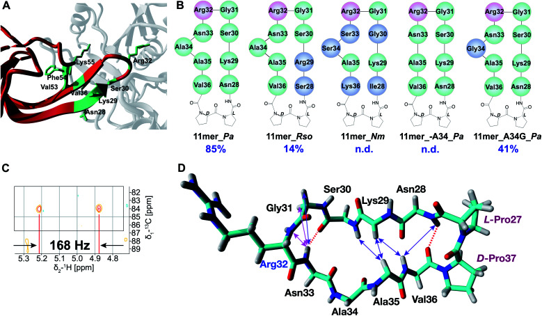
Fig. 2. (A) EarP–EF-P complex from P. aeruginosa (generated with YASARA, PDB 6J7M). EarP is depicted in grey, domain I of EF-P is colored red, EF-P residues involved in binding with EarP are in green. (B) Cyclic peptide mimics of the EF-P β-hairpin. Arg32 is shown in pink, altered residues (with respect to 11mer_Pa) are shown in blue. (C) Zoom-in of the coupled HSQC spectrum of crude Rha-11mer_Pa reaction mixture. JCH = 168 Hz (α-glycosidic bond). (D) Experimentally determined NOE signals that are indicative of a β-hairpin structure are mapped on the 11mer l-Pro–d-Pro_Pa fragment (from 3OYY crystal structure). Hydrogen bonds inferred from the NH temperature studies are shown as red dashed lines. NOE signals are shown as double-ended arrows (magenta: medium NOE; purple: weak NOE).

Fig. 2