. 2021 Jan 13;12(5):1620–1631. doi: 10.1039/d0sc06412b

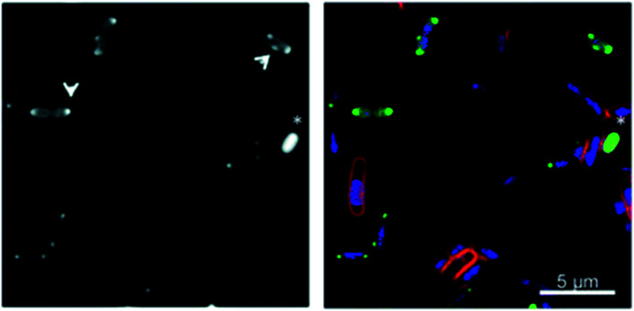
Fig. 5. Fluorescence microscopy of metallohelices in bacteria. Adapted from ref. 56 under licence CC BY 3.0, showing sub-cellular localization of Λ-9b′ in E. coli EHEC Sakai bacteria. Exponentially growing EHEC Sakai cells were treated with 8 μg mL−1 Λ-9b′ and stained for membrane (FM 4-64 FX), nucleic acid (DAPI) and the Λ-9b′ (via click reaction with AF-488 azide). (a) AF-488 staining (the arrows indicate the punctate localisation pattern of Λ-9b′, asterisk indicates an example of a bacterial cell with prominent Λ-9b′ but lack of DAPI staining). (b) is an overlay of membrane stain (red), nucleic acid staining (blue) and ‘click’ staining of Λ-9b′ (green).

Fig. 5