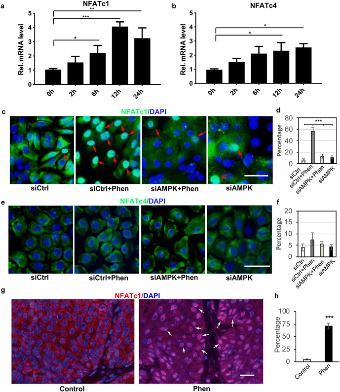
Figure 3. Phenformin promotes keratinocyte differentiation via the activation of CnB/NFAT signaling.
a-b: Human keratinocytes in suspension assays were collected at the indicated time points for qRT-PCR analysis of NFATc1 (a) and NFATc4 (b). c-f: Human keratinocytes transfected with siRNAs against AMPKα1/α2 (siAMPK) or a scrambled control siRNA (siCtrl), were cultured in the presence or absence of 1 mM phenformin (Phen), followed by IF analyses with anti-NFATc1 (green, c) or anti-NFATc4 antibody (green, e) together with DAPI staining for nuclei (blue). Red arrows indicate nuclear staining of NAFTc1. Percentages of nuclear positive staining of NFATc1 (d) or NFATc4 (f) in 200 cells were quantified from the images shown in c and e, respectively. g-h: Tumor sections from Figure 1 were used for IF analyses of NFATc1 (red) together with DAPI staining for nuclei (blue) (g) and were quantified (h). c, e: bar = 50 μm; g: bars = 100 μm. * P<0.05, **P<0.01, ***P<0.005. by Student’s two tailed t-test.

