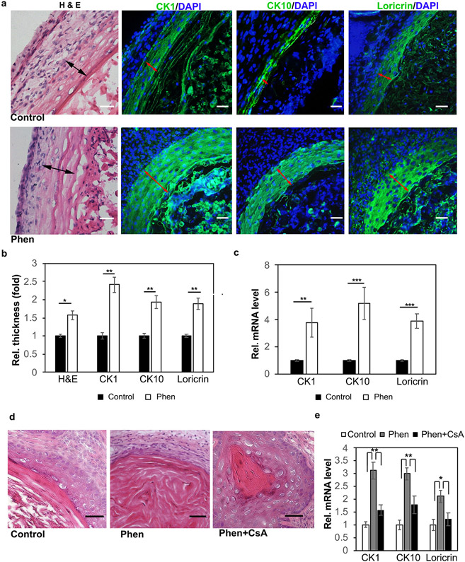
Figure 6. Phenformin induces human keratinocyte differentiation in vivo.
a: Human keratinocytes and dermal fibroblasts were injected into nu/nu mice, which were treated with phenformin (Phen) or with PBS (control). At 2 weeks, the grafts were collected for H&E stains, and IF staining of the differentiation markers CK1, CK10 and loricrin (green) as indicated. Black arrows indicate the keratinization zone of the epidermis in HE staining; red arrows indicate the positive staining zone of the epidermis. b: Quantification of area size indicated by black arrows in (a). c: Grafts collected from a were subjected to qRT-PCR analysis. d-e: Human skin cells prepared and grafted as described in (a); after grafting, mice were treated with drugs as indicated. At two weeks, the grafts were collected for H&E (d) or for RT-PCR analysis of differentiation markers (e). Bars = 100 μm. b, c, e: * P<0.05, **P<0.01, ***P<0.005. by Student’s two tailed t-test.

