. 2021 Jan 4;12(6):2138–2145. doi: 10.1039/d0sc03889j

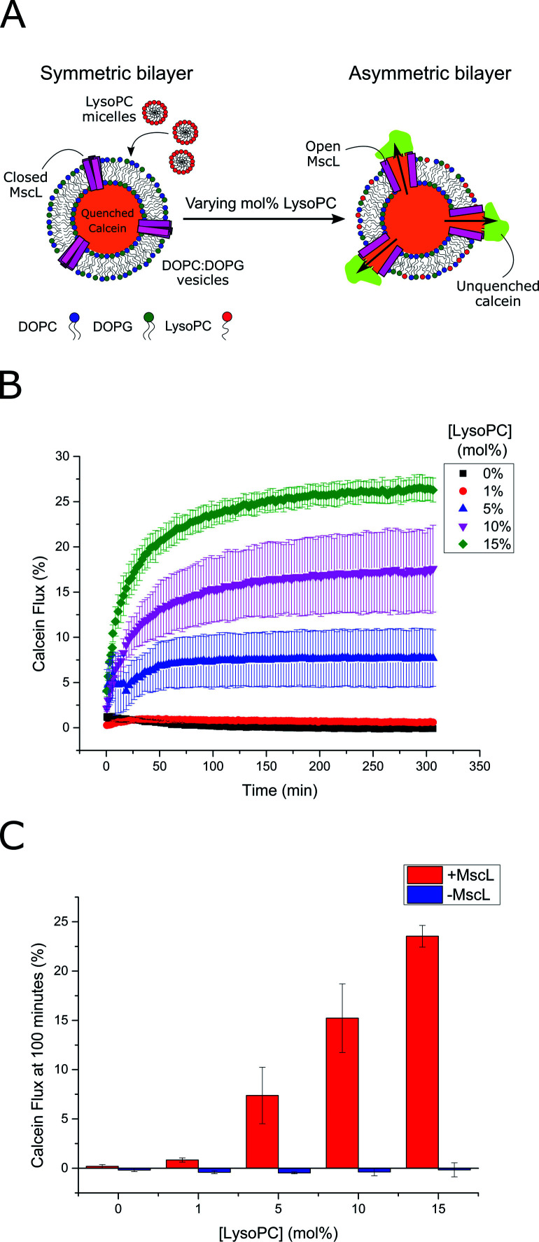
Fig. 2. LPC triggers MscL activation in lipid vesicles. (A) MscL is reconstituted into calcein-loaded lipid vesicles via detergent-mediated reconstitution. By encapsulating calcein at high concentrations, its fluorescence is fully quenched. Asymmetric insertion of LPC into the outer leaflet of the vesicle membrane is facilitated by the addition of LPC micelles to solution. If MscL is activated by LPC insertion, calcein will diffuse through the channel out of the vesicle where it will become diluted and generate a fluorescent signal as it becomes unquenched. (B) Calcein flux (%) over 5 hours from MscL-vesicles upon the addition of increasing mol% LPC to solution. LPC-dependent calcein flux can be observed indicating successful MscL activation by LPC. (C) MscL is critical for calcein flux. If calcein flux for vesicles ± MscL is assessed 100 minutes after LPC addition, negligible flux is observed at all LPC concentrations for vesicles lacking the channel. Error bars for (B) and (C) represent 1 SD (n = 3).

Fig. 2