. 2021 Jan 5;12(8):2970–2978. doi: 10.1039/d0sc05650b

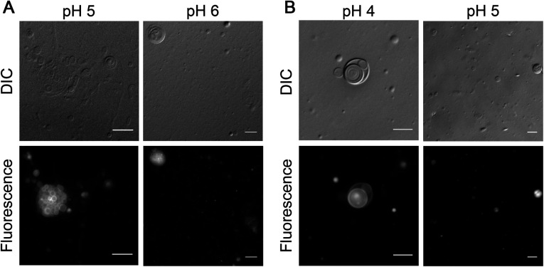
Fig. 6. Vesicle formation by NAAs under acidic conditions. (A) 6 mM of NOG forms vesicles in 200 mM acetate buffer at pH 5 (left panel) and in 200 mM MES buffer at pH 6 (right panel). (B) 6 mM NOS forms vesicles in 200 mM acetate buffer at pH 4 (left panel) and 5 (right panel). The observed vesicles typically vary in their size, shape and lamellarity with some being unilamellar, multilamellar and some even multivesicular in nature. Vesicle solutions were incubated at 60 °C for 1 hour and immediately subjected to microscopy. For better visualization, vesicles were stained with 10 μM of octadecyl rhodamine B chloride (R18) dye and observed using differential interference contrast (DIC) as well as fluorescence microscopy (see Methods for more details). Scale bar is 10 μm.

Fig. 6