. 2020 Jun 18;12(10):3379–3392. doi: 10.1039/d0sc02925d

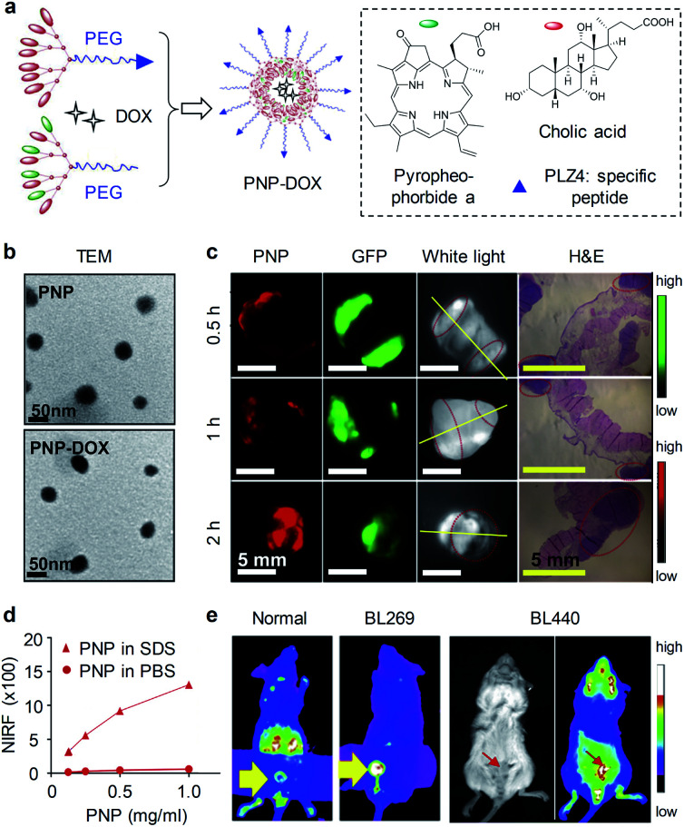
Fig. 12. (a) Schematic illustration of the preparation of PNP and PNP-DOX. The amino acid sequence of PLZ4: cQDGRMGFc. PEG has a molecular weight of 5000 Da. (b) TEM images of PNP and PNP-DOX. (c) PDD images in an MB49-GFP-Luc orthotopic bladder cancer mouse model after 2 h intravesical injection of PNPs (10 mg mL−1). Bladder was harvested and imaged in the GFP (tumor cells) and NIRF channel (PNP uptake) using Kodak imaging station. Yellow lines indicate cut line for histopathology; red circles indicate tumor locations. (d) NIRF intensity of different concentrations of PNPs in PBS and SDS. (e) Representative NIRF images of PNPs in orthotopic PDX BL269 and BL440 bladder cancer mice after intravesical administration of PNPs. Reproduced with permission from ref. 79. Copyright 2016, Elsevier.

Fig. 12