. 2020 Jun 18;12(10):3379–3392. doi: 10.1039/d0sc02925d

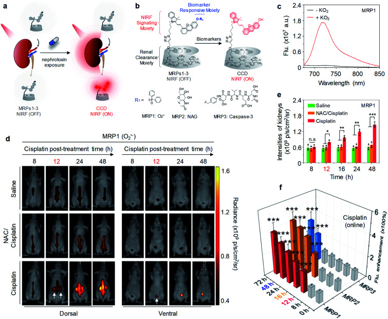
Fig. 5. (a) Schematic illustration of NIRF imaging of drug-induced AKI using MRPs1–3. (b) Chemical structures and molecular mechanisms of MRPs1–3 and their activated forms in response to O2˙, NAG and caspase-3, respectively, (R = H or CH2CHOHCH3). (c) Fluorescence spectra of MRP1 (30 μM) in the absence or presence of KO2 (60 μM) in PBS buffer (10 mM, pH 7.4). (d) Representative NIR fluorescence images of living mice with injection of MRP1 after treatment of cisplatin (20 mg per kg body weight) for 8, 12, 24 or 48 h. The control groups were treated with PBS or a nephroprotective antioxidant NAC. NIRF images acquired at 720 nm upon excitation at 675 nm. (e) NIRF intensities of kidneys in living mice after 30 min injection of MRP1 at the different post-treatment time points (8, 12, 16, 24 or 48 h). (f) Fluorescence enhancement of excreted MRPs1–3 in the urine from cisplatin-treated living mice after injection at different time points post-treatment of cisplatin. n.s: not significant, *p < 0.05, **p < 0.01, ***p < 0.001. Reproduced with permission from ref. 56. Copyright 2019 Springer Nature.

Fig. 5