. 2021 Jan 22;12(12):4345–4352. doi: 10.1039/d0sc05718e

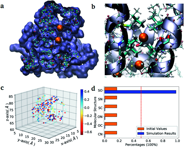
Fig. 3. Diatomic molecules in a protein environment. (a) X-ray crystal structure of the protein H-NOX (PBD entry code 3TFA) showing the main channel with three binding pockets occupied by Xe atoms (orange spheres). (b) Zoom into the first binding pocket with highlighted the apolar residues that favour the selection and diffusion of the gas molecules. The orange spheres indicate the position of the co-crystallized Xe atoms, which mark the center of the binding pockets. (c) Position of the point charges used to mimic the enzyme pocket. The red dot corresponds to the center of the diatomic molecules. (d) Initial and final distributions of the molecular structures obtained with the ‘alchemical’ optimization.

Fig. 3