. 2021 Feb 9;12(12):4300–4308. doi: 10.1039/d0sc06969h

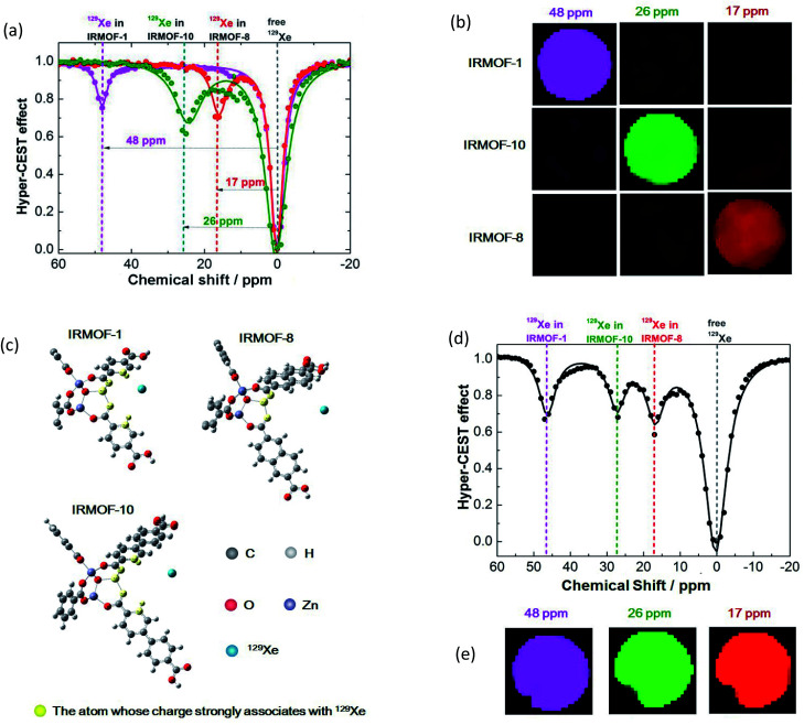
Fig. 2. Manipulation of the MOF pore structure to produce unique 129Xe MR signals. (a) Frequency-dependent saturation spectra of IRMOF-1, IRMOF-8, and IRMOF-10. To eliminate the influence of solvent, chemical shifts were referenced to the dissolved free 129Xe atom. (b) Coloring ultrasensitive MRI with three types of MOF nanoparticles. Each MOF is saturated at its unique chemical shift, thus enabling discrimination from others in ultrasensitive MRI. (c) Calculating the charge in an edge of an MOF pore. Charge numbers contribute to the differences in chemical shifts. (d) A mixture containing IRMOF-1, IRMOF-8, and IRMOF-10 showed separated signals. (e) MOF nanoparticles provide diverse contrast agents for complex mixture imaging. The MOF mixture showing ultrasensitive MRI in three colors, thus enabling the labeling of three different targets in a single sample.

Fig. 2