. 2021 Feb 25;12(15):5419–5429. doi: 10.1039/d0sc06760a

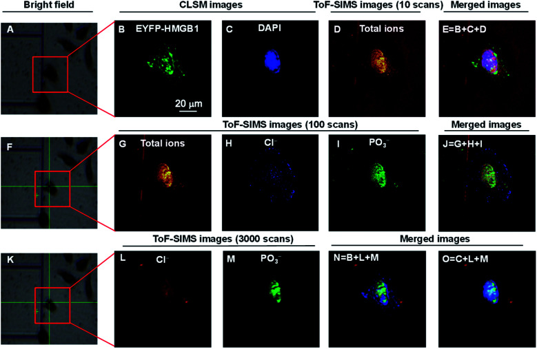
Fig. 2. COSIMSi of lyophilized HeLa cells without treatment using cisplatin. (A), (F) and (K) Bright field images of a single cell framed in a red box: (A) before; (F) after 10 cycles of GCIB sputtering on (A); and (K) after 100 BIB scans on (F). (B) and (C) CLSM images of (B) EYFP-HMGB1 and (C) the DAPI stained nucleus. (D) ToF-SIMS image of the total ions using 10 BIB scans. (E) Merged image of the CLSM images of EYFP-HMGB1 (green), DAPI (blue) and ToF-SIMS image of the total ions (red). (G)–(I) ToF-SIMS images of: (G) total ions; (H) Cl; and (I) PO3 using 100 BIB scans. (J) Merged image of the ToF-SIMS images of the total ions, Cl and PO3. (L) and (M) ToF-SIMS images of: (L) Cl; and (M) PO3 using 3000 BIB scans. (N) and (O) Merged ToF-SIMS images of Cl (red) and PO3 (green) and the CLSM image of EYFP-HMGB1 (blue) or DAPI (blue). The ToF-SIMS images of Cl and PO3 were acquired at m/z 35 and 79, respectively.

Fig. 2