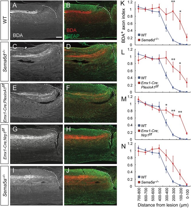
Figure 4.

Genetic deletion of Sema6d, PlexinA1, or Nrp1 suppresses dieback of CST fibers after SCI in mice. (A–J) Representative sagittal views of the thoracic spinal cord showing BDA-labeled CST axons in the rostral area of the lesion in WT (A, B), Sema6d−/− (C, D), Emx1-Cre; PlexinA1f/f (E, F), Emx1-Cre; Nrp1f/f (G, H), and Sema5a−/− mice (I, J) at day 56 post-injury. BDA, white (left panel) and red (right panel); GFAP, green. Dotted lines represent the rostral lesion borders. The arrowhead in (C) indicates defasciculated CST fibers. Scale bar, 200 μm. (K–N) Quantification of CST axon amounts in the bins rostral to the lesion site in WT (n = 5, blue), Sema6d−/− (K, n = 5), Emx1-Cre; PlexinA1f/f (L, n = 5), Emx1-Cre; Nrp1f/f (M, n = 6), and Sema5a−/− mice (N, n = 7). **P < 0.01, *P < 0.05, two-way repeated measures ANOVA and Tukey’s test.
