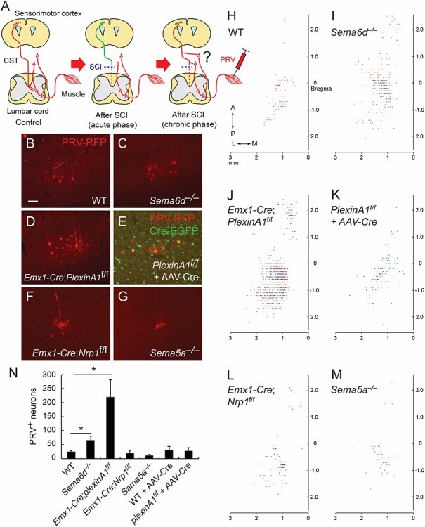
Figure 5.

Connectivity between the hindlimb muscle and cerebral cortex following SCI in semaphorin mutant mice. (A) Schema of the PRV tracing experiment from the hindlimb muscle to examine whether cortical neurons form new connections with spared circuits that bypass the lesion to the lower spinal levels (right panel). (B–G) Representative images of PRV-labeled layer V neurons (red) in WT (B), Sema6d−/− (C), Emx1-Cre; PlexinA1f/f (D), PlexinA1f/f + AAV-Cre-EGFP (E), Emx1-Cre; Nrp1f/f (F), and Sema5a−/− mice (G) after SCI at day 42 + 6. Scale bar, 50 μm. (H–M) Top views of the cortical locations of PRV+ layer V neurons in the cerebral cortices of WT (H), Sema6d−/− (I), Emx1-Cre; PlexinA1f/f (J), PlexinA1f/f + AAV-Cre (K), Emx1-Cre; Nrp1f/f (L), and Sema5a−/− mice (M) after SCI. Plots of three representative animals are shown in red, blue, and green. (N) The number of PRV+ layer V neurons in WT (n = 5), Sema6d−/− (n = 6), Emx1-Cre; PlexinA1f/f (n = 7), Emx1-Cre; Nrp1f/f (n = 5), Sema5a−/− (n = 6), WT + AAV-Cre (n = 6), and Emx1-Cre; PlexinA1f/f + AAV-Cre mice (n = 7). *P < 0.05, Mann–Whitney tests or unpaired t-tests.
