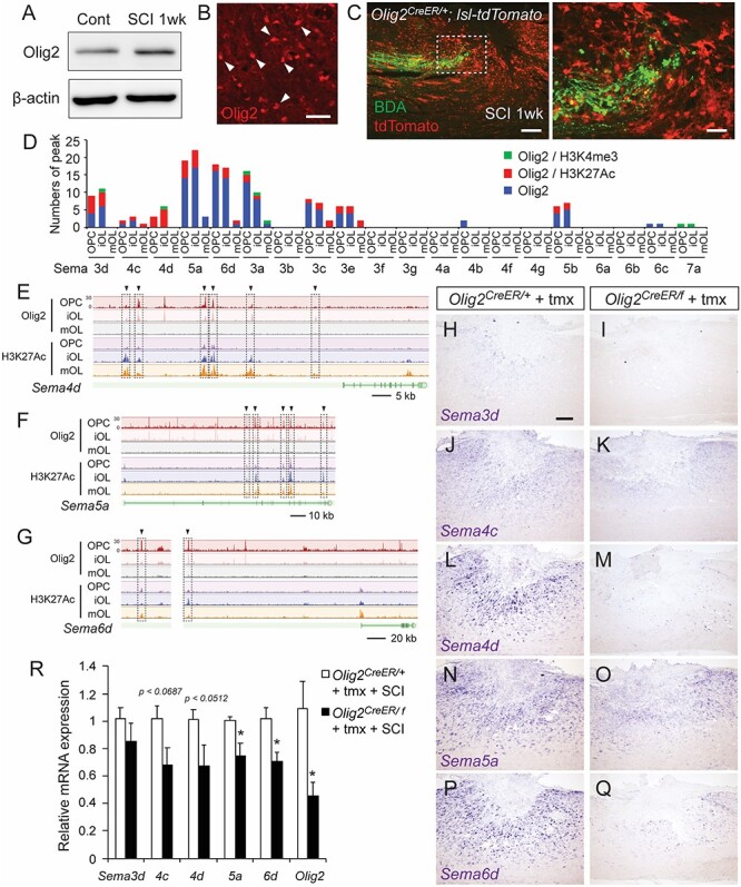
Figure 6.

Olig2 is required for semaphorin expression following SCI. (A) Western blots of the Olig2 protein in the thoracic cords of control and SCI mice at 1 week post-injury. (B) A number of Olig2+ cells were seen around the lesion. Scale bar, 25 μm. (C) Transected BDA+ CS axons (green) in front of accumulating Olig2+ cells labeled with tdTomato (red), in the rostral area of the lesion in Olig2CreER/+; lsl-tdTomato mice at day 7 post-injury. Sagittal section (right panel shows a magnified view of the dotted box). (D) The number of Olig2-binding peaks in genomic regions of Sema genes in ChIP-seq data acquired from OPCs, iOLs, and mOLs (Yu et al. 2013). Green, peaks corresponding to H3K4me3 binding peaks; red, peaks corresponding to H3K27Ac; blue, other Olig2 binding peaks. (E–G) ChIP-seq data of Olig2 and H3K27Ac in Sema4d (E), Sema5a (F), and Sema6d (G) genomic regions. Arrowheads and dotted squares represent the binding peaks common to Olig2 and H3K27Ac. (H–Q) In situ hybridizations of Sema3d (H, I), 4c (J, K), 4d (L, M), 5a (N, O), and 6d (P, Q) mRNA in the thoracic cords of tamoxifen-injected control Olig2CreER/+ and Olig2CreER/f mice at 1 week post-SCI. (R) Comparison of levels of Sema3d, 4c, 4d, 5a, and 6d mRNA expression in the thoracic cords of tamoxifen-injected Olig2CreER/+ and Olig2CreER/f mice at 1 week post-SCI, assessed by real-time PCR. N = 6, *P < 0.05, unpaired t-test. Scale bars, 25 μm (B), 200 μm (C, left), 50 μm (C, right), 200 μm (H–Q).
