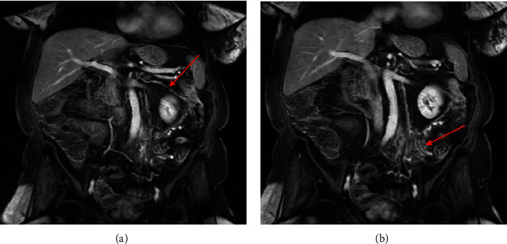
Figure 3.

MRA of the abdomen with and without gadolinium 7 weeks after initial presentation. Coronal T1-weighted fat-suppressed VIBE late arterial phase postcontrast images display (a) a prominent marginal artery connecting the middle colic artery to the left colic artery (arrow) and also reveal (b) a cluster of early draining veins in the region of the sigmoid colon mesentery (arrow).
