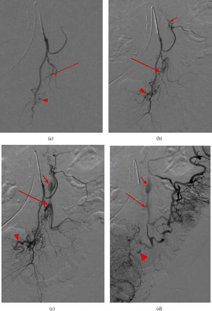
Figure 4.

Mesenteric angiogram demonstrates subselection of the IMA in order of progressive filling which eventually displays three AVM niduses arising from the left colic artery, sigmoid artery, and superior rectal artery with respective dilated venous outflow tracts. Minimal opacification is seen in the presumed venous outflow tracts (a) of the AVM niduses at the sigmoid (red arrow) and superior rectal artery (arrowhead). Progressive opacification (b) is noted in the sigmoid (long arrow) and superior rectal niduses (arrowhead); opacification of the left colic nidus is demonstrated (short arrow). Ectatic venous outflow tracts of the left colic (c) (short arrow), sigmoid (long arrow), and superior rectal (arrowhead) AVM niduses are seen. Washout of the venous outflow tracts of the three AVM niduses (d) is demonstrated with similar labeling as (b).
