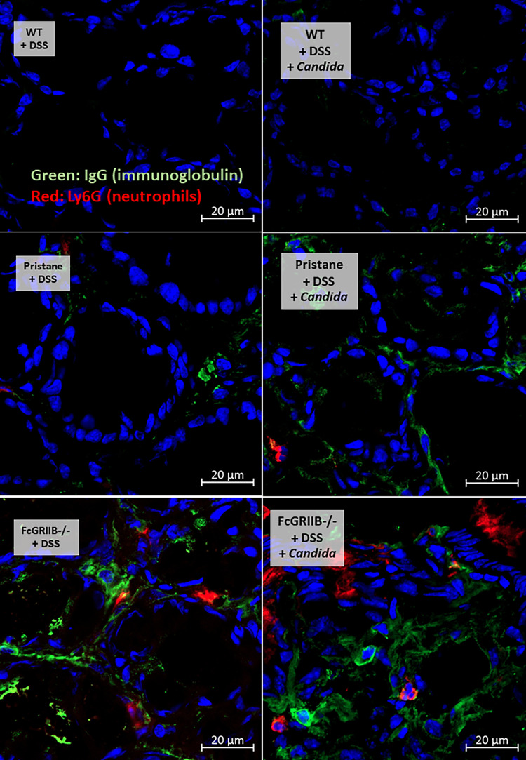
Figure 5.
Representative pictures of immunofluorescent stained sections for colon injury as determined by immunoglobulin G (IgG deposition) (green color of Alexa Fluor 488) and neutrophil accumulation (Ly6G) (red color of Alexa Fluor 647) of mice from wild-type (WT), Pristane and FcGRIIB-/- group after the administration of dextran sulfate solution (DSS) alone or with Candida gavage (DSS + Candida) (original magnification 630x) are demonstrated. Colon pictures from mice with control water in WT, Pristane and FcGRIIB-/- group are not presented due to the similarity to the represented pictures of WT+DSS and WT+DSS+Candida.

