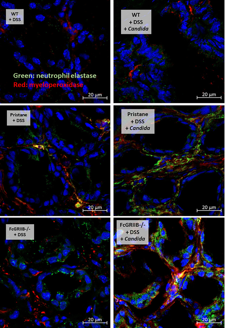
Figure 6.
Representative picture of immunofluorescent stained sections for neutrophil extracellular traps (NETs) in colons as determined by neutrophil elastase (NE) (green color of Alexa Fluor 488) and myeloperoxidase (MPO) (red color of Alexa Fluor 647) of mice from wild-type (WT), Pristane and FcGRIIB-/- groups after the administration of dextran sulfate solution (DSS) alone or with Candida gavage (DSS + Candida) (original magnification 630x) are demonstrated. Colon pictures from mice with control water in WT, Pristane and FcGRIIB-/- group are not presented due to the similarity to the represented pictures of WT+DSS.

