Figure 8.
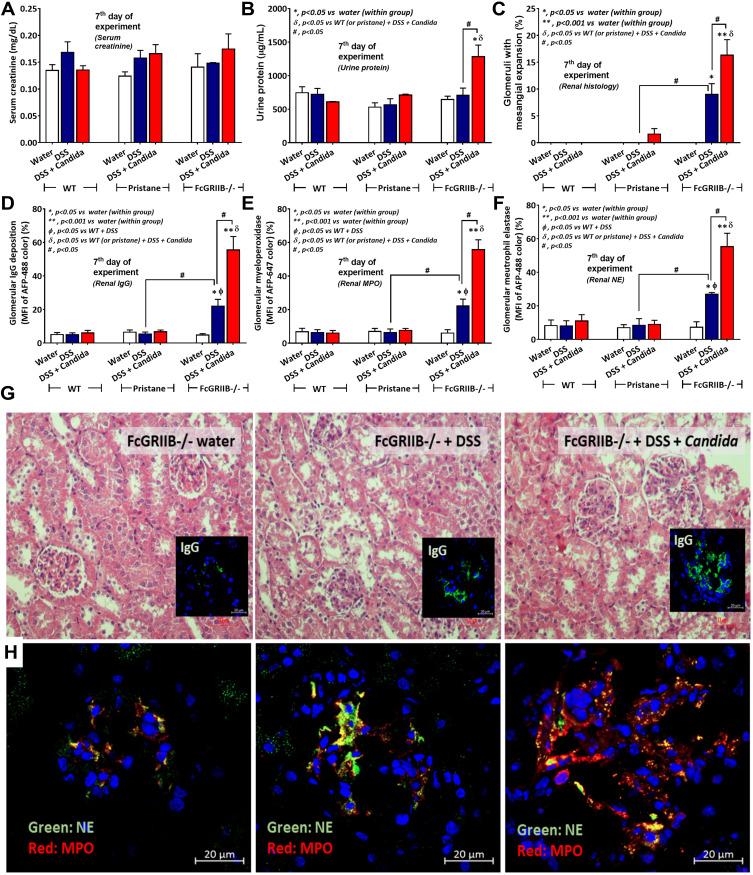
Characteristics of renal injury in FcGRIIB-/- mice after the administration of dextran sulfate solution (DSS) alone or with Candida gavage (DSS+Candida) as determined by serum creatinine (A), urine protein (B), glomerular injury (mesangial expansion) from the histology (C), immunoglobulin G (IgG) deposition in glomeruli (D) and glomerular neutrophil extracellular traps (NETs) as indicated by immunofluorescence of myeloperoxidase (MPO) (red color of Alexa Fluor 647) and neutrophil elastase (NE) (green color of Alexa Fluor 488) (E and F) are demonstrated (n = 6–9/group). Additionally, representative pictures of hematoxylin and eosin (H&E) stained section from kidney with immunofluorescence of glomerular IgG deposition (inset pictures) (G) and glomerular NETs formation (MPO and NE) (H) are demonstrated (original magnification 400x for H&E stain and 600x for immunofluorescence). Pictures from WT and Pristane mice are not presented due to the non-difference from FcGRIIB-/- mice with water control. *p < 0.05 vs water (within group); **p < 0.001 vs water (within group); ϕp < 0.05 vs WT+DSS; δp < 0.05 vs WT (or pristane) + DSS + Candida; #p < 0.05.

