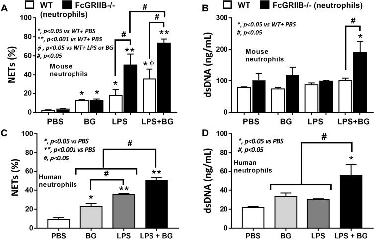
Figure 9.
Neutrophil extracellular traps (NETs) in mouse neutrophils, from wild-type (WT) and FcGRIIB-/- mice, and human neutrophils after the 2 h activation by phosphate buffer solution (PBS) or (1→3)-β-D-glucan (BG) or lipopolysaccharide (LPS) or LPS with BG (LPS+BG) are demonstrated. In mouse neutrophils, NETs were determined by the percentage of cells with NETs nucleus morphology using 4-,6-diamidino-2-phenylindole (DAPI), a nucleus stained color, with supernatant dsDNA by PicoGreen assay (A and B). In human neutrophils, NETs using the percentage of cells with positive staining for both myeloperoxidase (MPO) together with neutrophil elastase (NE) (merge color) (C) and the supernatant dsDNA (D) are demonstrated. All experiments were independently performed in triplicate. *p < 0.05 vs WT + PBS; **p < 0.001 vs WT + PBS; ϕp < 0.05 vs WT + LPS or BG; #p < 0.05.

