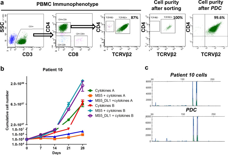
Fig. 1. Patient Sézary cells (SCs) in vitro expansion in defined media.
a Characterization of patient #10 SCs according to TCRVβ2, CD3, CD4, and CD8 expression. Dot plots representing the immunophenotype of PBMC, the purity of tumor TCRVβ2 + CD3 + CD4 + CD8-cells after flow cytometry cell sorting and the identification of SC purity after culture (patient-derived culture, PDC) by detection of TCRVβ2 + CD4+ cells using FACS. b Primary cultures of patient #10 SCs for 28 days after SC sorting. Cells were cultured on six different culture conditions and counted every week. c Identity of TCRγ gene rearrangement between original SCs of patient #10 and PDC was determined by the Biomed-2 protocol and capillary fragment analysis.

